Durr – VistaVox S Ceph

Durr – VistaVox S Ceph

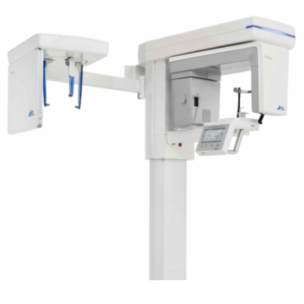
3D, 2D and Ceph X-ray images with exceptional image quality

The USP of the VistaVox S Ceph is found in the ideal 3D imaging volume which is oriented to the human anatomy. The jaw-shaped field of view of the VistaVox S Ceph maps the diagnostically relevant range of a 130-mm volume and is therefore visibly larger than the most commonly used volume of Ø 80 x 80 mm. The advantage: Thanks to this changed volume shape, VistaVox S Ceph also completely covers the region of the rear molars – an essential requirement for diagnostics, e.g. for an impacted wisdom tooth. In addition to that, VistaVox S Ceph offers ten further Ø 50 x 50 mm volumes: five each for the upper jaw and for the lower jaws. These are used if the indication only requires imaging of a certain region of the jaw, e.g. for endodontical or implantological treatments. Depending on the required level of detail in the image, the volumes can be used with a resolution of either 80 or 120 μm. Supplemented by the 17 panoramic programmes in the tried-and-tested S-pan technology, this provides dental practices with excellent imaging diagnostics in both the 2D and 3D areas.

Key features:

  • Ideal 3D imaging volume matched to the jaw arch (Ø 130 x 85 mm)
  • Ø 50 x 50 mm volumes in 80 or 120 μm resolution
  • Excellent image quality in 2D and 3D thanks to the high-resolution CsI sensor with a pixel size of 49.5 μm
  • Reduced radiation dose thanks to the anatomically adapted volume
  • VistaSoft – modern, ergonomic image processing software

Description

2D images with exceptional image quality

VistaVox S Ceph offers not only excellent value for money, but will also help you and your surgery team to increase your flexibility. In addition to CBCT images, the S-pan technology of the VistaVox S Ceph can be used to generate brilliant panoramic images, which set new standards in the image sharpness of extraoral images.

Key features:

  • S-Pan technology for easier diagnostics
  • CsI sensor for improved image quality and reduced radiation exposure
  • Extremely fast, panoramic images in 7 sec.
  • Tolerant of typical positioning errors – thanks to the S-Pan technology

Time-saving Cephalometric exposure with a low X-ray dose

Short scan time and high image quality with a low X-ray dose

The very short scan time of just 1.9 seconds helps to avoid motion artefacts and to reduce the radiation dose. The modern high-sensitivity CsI sensors enable excellent image quality.

3-in-1 X-ray system

In addition to the various CBCT volumes and the 17 panoramic programs, VistaVox S Ceph also offers six modes for all types of cephalometric exposures:

  • Head Lateral
  • Head Full Lateral
  • Head PA
  • SMV (submentovertex)
  • Waters View
  • Hand

Saving you time and money

VistaVox S Ceph is equipped with two high-end CsI sensors. The advantage: there is no need for the cumbersome process of unplugging and reconnecting between the 3D X-ray unit and the Ceph boom. To start a Ceph X-ray image acquisition, all you need to do is select the corresponding program mode.

Technical dataVistaVox S CephX-ray HV generator
Voltage, current60–99 kV, 4–16 mATube
Focal point0.5 mm (IEC60336)Total filtration2.8 mm ALImage detector
TypeCsI CMOS Photodiode arrayPixel size49.5 μm / 100 µmActive sensor surface135.8 x 36.4 mm / 157.2 x 16.3 mmScan times
Scan timesFrom 2 to 18 seconds for lateral head imagesCeph programmesin quick scan mode: 1.9 seconds (line scan)3D volumeØ 130 x 70 mm diagnostic
Ø 130 x 85 mm diagnostic
Ø 50 x 50 mmUnit dimensions
Height1,406–2,206 mmWeight202 kgHeight adjustment range800 mmWidth x depth1,540 x 1,190 mmInstallationWall mountingElectrical connections
Mains voltage200–240 V ACFrequency50/60 HzRated power2.2 kVA

See our other Durr Dental Products

Title

Go to Top