Description
All functions at a glance
The intuitive 7″ Touch-Display visualises all the settings; quickly and in high resolution. The user- friendly handling and navigation produce an exceptionally smooth X-ray procedure. Simply select the X-ray program and the patient size to prevent handling errors, thereby ensuring an optimal workflow.
Always the right image size
With an image height of 150 mm, the VistaPano S displays both an extended diagnosable jaw area, whilst producing a high-quality image of the paranasal sinuses. Selection of the the child program significantly reduces radiation exposure through the use of a lower collimator and a shortened scanning time.
Panorama X-ray programs
With a total of 17 X-ray programs, you are well equipped for every diagnostic requirement. In addition to the standard panorama program, VistaPano S offers:
▪ Half-side recordings of right, left and front
▪ 4 child programs: a recording mode with smaller exposure area; a 45 – 56 % reduction in the dose without a concurrent loss of diagnostic information
▪ 5 programs for orthogonal X-ray images
▪ 2 programs for temporomandibular imaging
(functional diagnosis)
▪ 2 programs for sinus X-ray images to display paranasal sinuses
S-Pan technology: High resolution images produce safe diagnosis
Conventional digital devices generate panoramic images along a prescribed contour following the jaw line of an
‘average’ person on the axial plane. They reproduce a layer like an unrolling piece of level paper placed along this contour. This usually involves a compromise.
S-Pan technology works entirely differently. The image sections are automatically selected from a range of parallel layers, which best correspond to the patient anatomy. These image parts are placed together to form a panoramic image, which focuses on patient anatomy, taking into account unusually- shaped dentition and individually-angled teeth. The result is an image of impressive clarity. The dentist is able to locate immediately all the structures of interest, without any further intervention. As the reconstruction is oriented to the actual position of the bite, incorrect positioning is balanced out to a certain extent. This saves time and repetition.
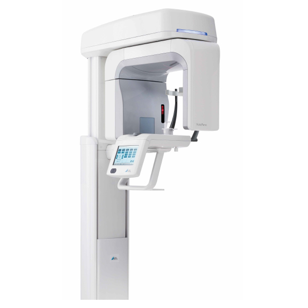
Fits in every dental practice
The streamlined, delicate design of the new VistaPano S Ceph permits a range of placements in your surgery rooms. Its design makes it an attractive eye-catcher in any location. Its dimensions of 1.0 x 2.3 x 1.5 m (W x D x H) requires the smallest of space.
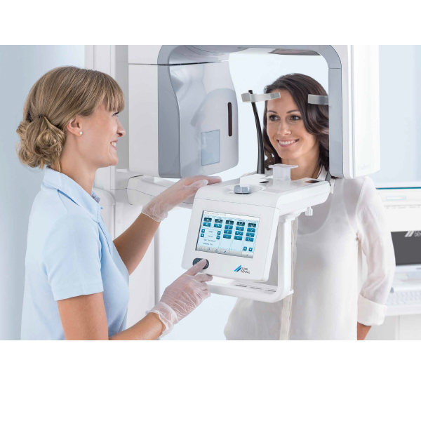
Simple and efficient patient positioning
Three laser guide lines mark the exact orientation of Frankfurt horizontal plane, median sagittal plane, and film plane. The face-to-face positioning without a mirror allows direct visual contact between operator and patient, thus promoting the sense of well-being and hence the co-operation of the patient.
| Technical Data |
VistaPano S |
| X-ray HV generator |
|
| Voltage, current |
50 – 99 kV, 4 – 16 mA |
| Tubes |
|
| Focal spot |
0.5 mm (IEC60336) |
| Total filtration |
2.8 mm Al eq. |
| Image detector |
|
| Model |
CsI sensor |
| Pixel size |
100 μm |
| Active sensor size |
6 x 150.4 mm |
| Frame rate |
300 fps |
| Scan times |
|
| Scan times |
From 2.5 to 13.5 secs. |
| Panorama program scan times |
Panoramic X-ray image of adults in quick scan mode: 7 sec. |
| Magnification factor |
|
| Magnification factor |
1.3 |
| |
|
| Device dimensions |
|
| Maximum height |
2280 mm |
| Weight (without/with foot) |
105/155 kg |
| Height adjustment range |
700 mm |
| Width x depth x height |
990 x 1220 x 2280 mm |
| Installation |
Wall mounting or foot |
| Electrical connection |
|
| Mains voltage |
200 – 240 V AC |
| Frequency |
50/60 Hz |
| Rated power |
2.2 kVA |