3D Dental CBCT

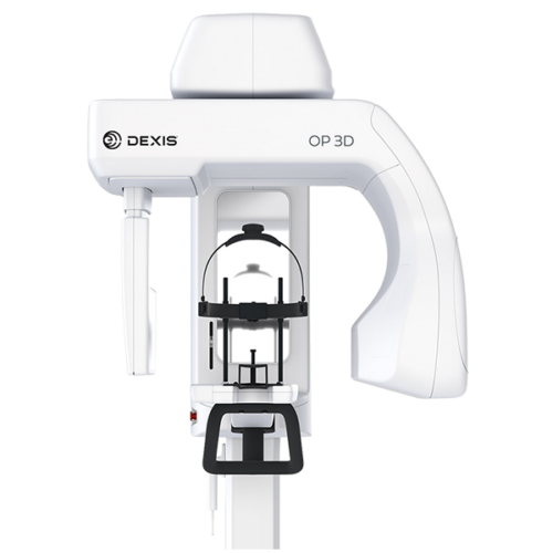
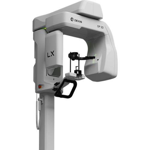
  • Simplify, streamline, and expand your diagnostic capabilities The next generation of proven DEXIS cone beam technology

    Built on OP 3D technology, this multimodality imaging platform expands your 3D diagnostic capabilities with a wide range of clinical applications that support your evolving practice and enhance diagnostic confidence. The 2D and 3D imaging options cover a full spectrum of dental extraoral needs, from endodontics to complex surgical cases. This next-generation system offers flexible FOV* options ranging from 5x5 up to 15x20 – the largest view option available on a DEXIS OP 3D platform to date. With shorter scan times, the OP 3D LX captures the maxillofacial complex and large diagnostic areas in one non-stitched scan for fast workflows.

  • ORTHOPANTOMOGRAPHTM™ OP 3D is a powerful diagnostic tool for 3D imaging, panoramic, and cephalometric imaging. The OP 3D makes choosing your X-ray system simple. It is a complete X-ray platform that provides easy-to-use features throughout the entire dental imaging workflow.

    With its versatile imaging programs and intuitive user interface, the DEXIS OP 3D in its different configurations offers imaging excellence for a variety of users, ranging from general dental practitioners to orthodontists, all the way to maxillofacial surgeons.

    DTX Studio™ suite connects the devices and technologies in your dental practice or lab – in one single platform.

     
  • IT'S TIME FOR EFFICIENT 3D DIAGNOSIS

    X-Mind® prime 3D is the latest addition to ACTEON’s CBCT digital imaging product line. It blends cutting-edge technology with ease of use in a compact design. With X-Mind® prime 3D, high tech is at your fingertips with tools for accurate diagnosis and complete treatment planning. X-Mind® prime 3D is a lightweight and compact system that can fit into virtually any dental office. With its intelligent, wall-mounted design, the unit takes up zero floor space. Because the unit is delivered preassembled to the office, it can be installed by a single technician in under an hour, so it is up and running and does not interfere with office production. 3D DIAGNOSIS IS MORE ACCESSIBLE THAN EVER WITH A SMART AND COMPACT SOLUTION
  • 3D, 2D and Ceph X-ray images with exceptional image quality

    The USP of the VistaVox S Ceph is found in the ideal 3D imaging volume which is oriented to the human anatomy. The jaw-shaped field of view of the VistaVox S Ceph maps the diagnostically relevant range of a 130-mm volume and is therefore visibly larger than the most commonly used volume of Ø 80 x 80 mm. The advantage: Thanks to this changed volume shape, VistaVox S Ceph also completely covers the region of the rear molars – an essential requirement for diagnostics, e.g. for an impacted wisdom tooth. In addition to that, VistaVox S Ceph offers ten further Ø 50 x 50 mm volumes: five each for the upper jaw and for the lower jaws. These are used if the indication only requires imaging of a certain region of the jaw, e.g. for endodontical or implantological treatments. Depending on the required level of detail in the image, the volumes can be used with a resolution of either 80 or 120 μm. Supplemented by the 17 panoramic programmes in the tried-and-tested S-pan technology, this provides dental practices with excellent imaging diagnostics in both the 2D and 3D areas.

    Key features:

    • Ideal 3D imaging volume matched to the jaw arch (Ø 130 x 85 mm)
    • Ø 50 x 50 mm volumes in 80 or 120 μm resolution
    • Excellent image quality in 2D and 3D thanks to the high-resolution CsI sensor with a pixel size of 49.5 μm
    • Reduced radiation dose thanks to the anatomically adapted volume
    • VistaSoft – modern, ergonomic image processing software
  • Taking 3D diagnostics to the next level - VistaVox S generates a 3D volume ideally adapted to the contour of the jaw

    The USP of the VistaVox S is found in the ideal 3D imaging volume which is oriented to the human anatomy. The jaw-shaped field of view of the VistaVox S maps the diagnostically relevant range of a 130-mm volume and is therefore visibly larger than the most commonly used volume of Ø 80 x 80 mm. The advantage: Thanks to this changed volume shape, VistaVox S also completely covers the region of the rear molars – an essential requirement for diagnostics, e.g. for an impacted wisdom tooth. In addition to that, VistaVox S offers ten further Ø 50 x 50 mm volumes: five each for the upper jaw and for the lower jaws. These are used if the indication only requires imaging of a certain region of the jaw, e.g. for endodontical or implantological treatments. Depending on the required level of detail in the image, the volumes can be used with a resolution of either 80 or 120 μm. Supplemented by the 17 panoramic programmes in the tried-and-tested S-pan technology, this provides dental practices with excellent imaging diagnostics in both the 2D and 3D areas.

    Key features:

    • Ideal 3D imaging volume matched to the jaw arch (Ø 130 x 85 mm)
    • Ø 50 x 50 mm volumes in 80 or 120 μm resolution
    • Excellent image quality in 2D and 3D thanks to the high-resolution CsI sensor with a pixel size of 49.5 μm
    • Reduced radiation dose thanks to the anatomically adapted volume
    • VistaSoft – modern, ergonomic image processing software

    2D images with exceptional image quality

    VistaVox S offers not only excellent value for money, but will also help you and your surgery team to increase your flexibility. In addition to CBCT images, the S-pan technology of the VistaVox S can be used to generate brilliant panoramic images, which set new standards in the image sharpness of extraoral images.

    This is what counts:

    • S-Pan technology for easier diagnostics
    • CsI sensor for improved image quality and reduced radiation exposure
    • Extremely fast, panoramic image in 7 sec.
    • Tolerant of typical positioning errors – thanks to the S-Pan technology
  • The First Line of Upgradeable Dedicated 3D CBCT Machines

    This dynamic system offers three maximum fields of view (FOV), V8, V10, and V17. Within these sizes, you also have the option to scale or collimate the scan height to capture only the area of interest per the patient’s immediate need. The system also provides enhanced low-dose imaging options through protocols and collimation, dedicated traditional 2D panoramic capabilities, and the feature-rich Tx STUDIO™ software, powered by Anatomage—all at a price point starting at under $90,000. i-CAT™ FLX V8 – With 8 cm x 5 cm to 8 cm x 8 cm FOVs, this system accommodates those dentists who want to capture select treatment areas for survey, implant, oral surgery and endodontics. i-CAT FLX V10 – The V10 offers a maximum and scalable FOV of up to 16 cm D x 10 cm H, helpful when you expand into larger cases and airway or TMJ analysis. i-CAT FLX V17 – With a maximum FOV of up to 23 cm D x 17 cm H, the V17 is used most often by orthodontics, oral maxillofacial surgeons and oral radiologists.

  • The Most Comprehensive 3-in-1 for the Entire Maxillofacial Region

    The ORTHOPANTOMOGRAPH™ OP 3D Pro, by DEXIS™, crystallizes the 50-year heritage of a legendary product family. It uses the latest in 3D and 2D technologies, for your benefit. The modern 3-in-1 platform’s flawless design and versatility set the benchmark for whole maxillofacial imaging.
    • 3-in-1 platform delivers high-quality images
    • Versatile selection of volumes and resolutions
    • Automatic dose control (ADC) technology with 3D
    • SMARTVIEW™ feature for accurate FOV positioning
    • Free FOV positioning in horizontal and vertical directions
    • Metal Artifact Reduction (MAR) tool
    • Large, easy-to-use 10-inch user interface enables efficient usage
    • Intuitive patient positioning
    • Designed to be upgradeable and expandable with a cephalometric option available
     
  • Award winning innovations for panoramic, cephalometric and 3D imaging The DEXIS OP 3D makes choosing your X-ray system simple. It is a complete X-ray platform that provides easy-to-use features throughout the entire dental imaging workflow. With its versatile imaging programs and intuitive user interface, the DEXIS OP 3D in its different configurations offers imaging excellence for a variety of users, ranging from general dental practitioners to orthodontists, and all the way to maxillofacial surgeons. DTX Studio™ suite connects the devices and technologies in your dental practice or lab – in one single platform. The DEXIS OP 3D has won the German Innovation Award in 2018, which represents an externally recognised sign of innovation and quality. The DEXIS OP 3D has won the German Innovation Award in 2018, which represents an externally recognised sign of innovation and quality.

Title

Go to Top